Ultrasound (USG): A Comprehensive Diagnostic Imaging Tool
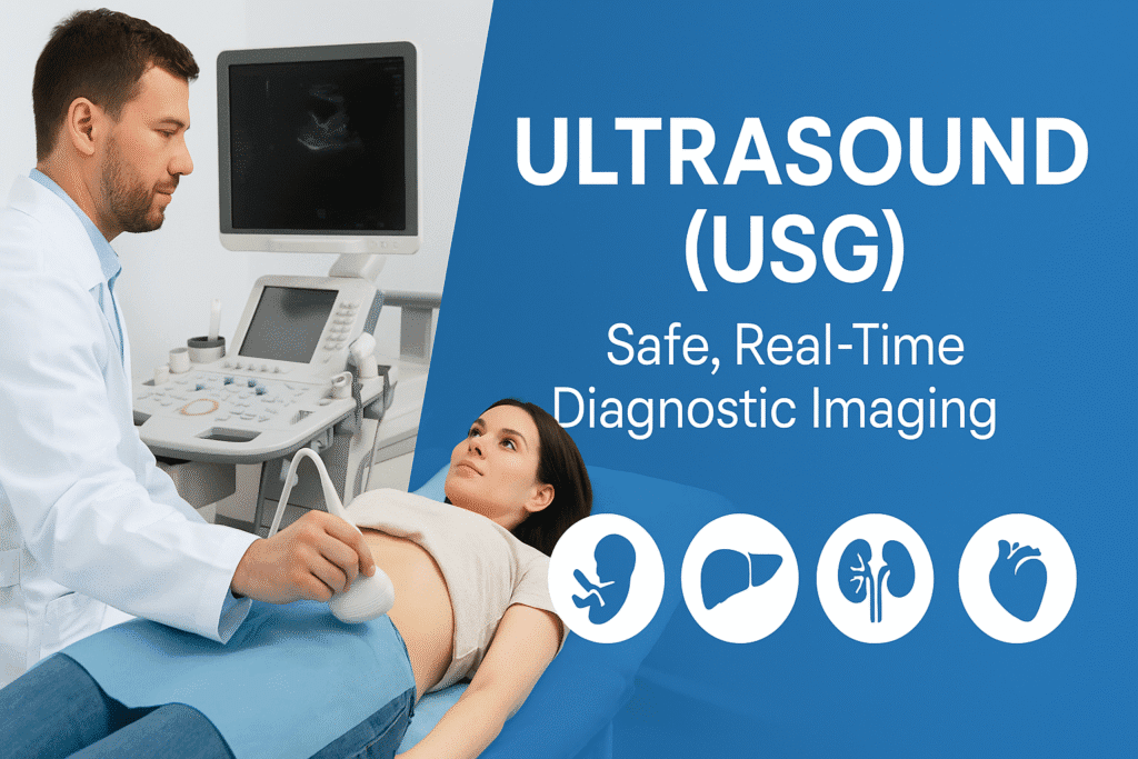
Ultrasound, also known as Ultrasonography (USG), is a non-invasive imaging technique that uses high-frequency sound waves to produce real-time images of internal body structures. It is widely used across various medical disciplines for diagnostic and therapeutic purposes. Its safety, accessibility, and lack of radiation make it an indispensable tool in modern medicine, especially in obstetrics, gynecology, abdominal imaging, urology, cardiology, and musculoskeletal medicine.
1. What is Ultrasound (USG)?
Ultrasound involves the use of a transducer (probe) that emits high-frequency sound waves into the body. These waves bounce off internal structures and return echoes, which are captured by the probe and processed by a computer to form live images on a screen.
Key Features:
-
Real-time imaging
-
Non-ionizing radiation (unlike X-rays or CT scans)
-
Portable and affordable
-
Safe for pregnant women and children
2. Types of Ultrasound
| Type | Purpose |
|---|---|
| Transabdominal USG | Common for abdominal, pelvic, and pregnancy scans |
| Transvaginal USG | Detailed pelvic imaging in females |
| Transrectal USG | Prostate imaging in males |
| Obstetric USG | Fetal monitoring during pregnancy |
| Doppler USG | Blood flow studies |
| Echocardiography | Heart structure and function |
| Musculoskeletal USG | Tendons, muscles, joints imaging |
| Endoscopic Ultrasound (EUS) | GI tract and pancreas imaging |
| 3D/4D Ultrasound | Advanced fetal and anatomical imaging |
3. Preparation Before Ultrasound
Preparation varies depending on the type of USG being performed.
General Preparations:
| USG Type | Patient Preparation |
|---|---|
| Abdominal USG | Fasting for 6–8 hours (to reduce bowel gas and allow better liver/gallbladder visualization) |
| Pelvic USG | Full bladder required—drink 3-4 glasses of water 1 hour prior, avoid urination |
| Pregnancy USG (early) | Full bladder improves visibility |
| Transvaginal USG | Empty bladder required |
| Renal/KUB USG | Hydration before scan; avoid urination if bladder needs to be visualized |
| Doppler USG | No specific preparation unless advised |
| Echocardiography | No fasting required |
4. Procedure of Ultrasound
-
Patient Positioning: The patient lies on an examination table; position depends on the organ to be visualized.
-
Application of Gel: A water-based gel is applied to the skin over the area being examined. This eliminates air pockets and improves sound wave transmission.
-
Probe Movement: The technician or radiologist moves the transducer across the skin. In transvaginal or transrectal USG, a covered and lubricated probe is gently inserted.
-
Image Acquisition: Images are seen in real-time. The radiologist may capture still images or videos for documentation.
-
Duration: Usually 15–30 minutes depending on complexity.
-
Post-scan: Gel is wiped off, and normal activities can be resumed immediately.
5. Clinical Importance of Ultrasound
A. In Obstetrics and Pregnancy Monitoring
Ultrasound plays a crucial role throughout pregnancy, from conception to delivery. Major types of pregnancy scans include:
| Scan | Timing | Purpose |
|---|---|---|
| Dating Scan (Viability scan) | 6–9 weeks | Confirm pregnancy, gestational age, detect heartbeat |
| NT Scan (Nuchal Translucency) | 11–13+6 weeks | Screen for Down Syndrome and chromosomal abnormalities |
| Anomaly Scan (Level II scan) | 18–22 weeks | Detailed fetal anatomy check, detect structural defects |
| Growth Scan | 28–32 weeks | Monitor fetal growth, amniotic fluid, placenta |
| Biophysical Profile (BPP) | After 32 weeks | Evaluate fetal well-being (movement, tone, breathing, AFI) |
| Doppler Scan | Any time after 28 weeks | Assess blood flow in umbilical artery and fetal vessels |
| Transvaginal Scan (TVS) | Early pregnancy or cervical length measurement |
Ultrasound in pregnancy helps in:
-
Detecting ectopic pregnancy
-
Identifying multiple gestations
-
Monitoring placental location (e.g., placenta previa)
-
Estimating fetal weight
-
Guiding amniocentesis and other invasive tests
✅ Note: Ultrasound is considered completely safe during pregnancy when used judiciously.
B. Abdominal and Pelvic Imaging
Used for:
-
Liver, gallbladder, pancreas, spleen, kidneys
-
Ascites (fluid in abdomen)
-
Abdominal aortic aneurysm
-
Urinary bladder volume and prostate size
-
Ovarian cysts, uterine fibroids, endometrial thickness
C. Cardiac Evaluation (Echocardiography)
-
Evaluates heart chambers, valves, wall motion
-
Essential in diagnosing heart failure, valvular disease, congenital heart disease
D. Doppler Ultrasound
-
Measures blood flow velocity in arteries and veins
-
Used in:
-
Deep vein thrombosis (DVT)
-
Varicose veins
-
Carotid artery stenosis
-
Umbilical artery in high-risk pregnancies
-
E. Emergency and Interventional Use
-
Trauma (FAST – Focused Assessment with Sonography for Trauma)
-
Guided procedures: aspiration, biopsy, catheter placement
-
Paracentesis, thoracentesis
6. Advantages of Ultrasound
-
No radiation exposure
-
Painless and non-invasive
-
Real-time guidance for interventions
-
Widely accessible and cost-effective
-
Portable, useful in bedside and rural settings
7. Limitations of Ultrasound
-
Limited by obesity or bowel gas
-
Operator-dependent
-
Cannot penetrate bone or air-filled structures (e.g., lungs, intestines)
-
Not as detailed as CT or MRI for some pathologies
8. Advanced Ultrasound Technologies
-
3D/4D Ultrasound: Detailed anatomical imaging; commonly used in fetal imaging
-
Elastography: Assesses tissue stiffness (e.g., in liver fibrosis)
-
Contrast-Enhanced Ultrasound (CEUS): Better lesion characterization
-
Endoscopic Ultrasound (EUS): High-resolution imaging of pancreas, GI tract
Conclusion
Ultrasound (USG) is a versatile, safe, and essential diagnostic tool in both routine and emergency settings. From monitoring pregnancy to diagnosing abdominal conditions and vascular abnormalities, its applications are vast. Patients should follow specific preparation instructions for accurate results. As technology evolves, newer applications like elastography and contrast-enhanced USG are expanding its diagnostic capabilities even further.
✅ FAQs on Ultrasound (USG)
1. Is ultrasound safe during pregnancy?
Yes, ultrasound is completely safe during pregnancy as it does not use ionizing radiation.
2. Can I eat before an abdominal ultrasound?
No, you are usually advised to fast for 6–8 hours before an abdominal USG for better image clarity.
3. What is the difference between transabdominal and transvaginal ultrasound?
Transabdominal is done externally on the belly, while transvaginal uses an internal probe for clearer pelvic organ images.
4. How long does a typical ultrasound take?
Most ultrasounds take between 15 to 30 minutes, depending on the body part and complexity.
5. What are Doppler ultrasounds used for?
Doppler studies blood flow in arteries and veins, often used in pregnancy, vascular diseases, and DVT diagnosis.
6. Is there any pain involved in ultrasound?
Ultrasound is painless. However, transvaginal or transrectal procedures may cause mild discomfort.
7. How soon will I get my ultrasound report?
Reports are usually available within a few hours to a day, depending on the facility and urgency.
For more regular updates you can visit our social media accounts,
Instagram: Follow us
Facebook: Follow us
WhatsApp: Join us
Telegram: Join us