All living organisms, including microorganisms, are classified as either prokaryotes or eukaryotes. Prokaryotes and eukaryotes are distinguished by their cellular characteristics. Prokaryotic cells lack nuclei and organelles, whereas eukaryotic cells contain both. Prokaryotic and eukaryotic cells share a number of characteristics. Both kinds of cells use DNA to store their genetic information and are surrounded by cell membranes, also known as plasma membranes. Prokaryotes are various types of microorganisms, including bacteria and cyanobacteria. Eukaryotes include microorganisms like fungi, protozoa, and simple algae. Viruses are not classified as prokaryotes or eukaryotes because they lack the characteristics of living things, with the exception of the ability to replicate (which they achieve only in living cells).
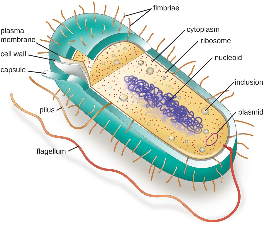
Prokaryotic cells
Prokaryotic cells are primitive types of cells that lack a nucleus. Furthermore, prokaryotes lack membrane-bound cellular organelles. Prokaryotes are exclusively unicellular. Prokaryotic cells are characterized by their ability to contain bacteria, cyanobacteria (formerly referred to as blue-green algae), rickettsiae, chlamydiae, and mycoplasmas.
Size and shapes
Prokaryotes, which range in size from 0.15 μm (mycoplasmas) to 0.25 μm (chlamydiae) to 0.45 μm (rickettsiae) to roughly 2.0 μm (many bacteria), are most likely the smallest living organisms. Certain prokaryotes, such as bacteria, occur in spherical forms called cocci (singular, coccus) or in rodlike forms called bacilli (singular, bacillus). Some bacteria have a comma shape (vibrio), or a flexible, wavy shape (spirochete), or a corkscrew shape (spirillum).
Some prokaryotes have a variety of shapes and sizes and are said to be pleomorphic. Rickettsiae and mycoplasmas are examples of pleomorphic microorganisms.
Some prokaryotes adhere to one another in a unique way when they split. For instance, a diplococcus is made up of two cocci, a streptococcus is made up of a chain of cocci, and a tetracoccus is made up of four cocci arranged in the shape of a cube. A staphylococcus is a cluster of cocci that resembles grapes. Streptobacilli are long chains of bacteria that can occasionally form.
Cell wall and cell membrane
Every bacterium, except mycoplasmas, has a semirigid cell wall. Since materials in the cytoplasm exert osmotic pressures, the cell wall gives the organisms shape and keeps them from bursting.
Prokaryotic cell walls primarily consist of peptidoglycan, a large polymer made up of N-acetylglucosamine and N-acetylmuramic acid. Gram-positive bacteria’s cell walls contain more peptidoglycan, which may explain their ability to retain the stain during the Gram stain procedure. Gram-negative bacteria contain more lipids in their cell walls. Teichoic acid polymers are commonly found in Gram-positive bacteria’s peptidoglycan.
Gram-negative bacteria have an outer membrane, a very thin layer that surrounds them in addition to their cell wall. Endotoxins are lipopolysaccharides that are a component of this outer membrane. The cell wall and outer membrane are separated by an area known as the periplasmic space, which is home to a material known as periplasm.
The cytoplasm of all prokaryotes is encased in a membrane called the plasma membrane, which is the cell membrane. Because the cell membrane follows the fluid mosaic model, its proteins are suspended in a double layer of phospholipids. Prokaryotes have respiratory enzymes at their cell membranes, which also have attachment sites for bacterial flagella and aid in DNA replication.
Cytoplasm
Prokaryotic cells’ cytoplasm is home to ribosomes and other granules that the organism uses. The DNA is contained in the nuclear region (the nucleoid) and has no histone protein to support it. Prokaryotic cells have in their cytoplasm a single, looped chromosome, as well as numerous small loops of DNA called plasmids. It appears that the plasmids’ genetic material is not necessary for the organism to survive in the long run.
Protein synthesis takes place in prokaryotic ribosomes, which also contain ribonucleic acid (RNA) and proteins. 70S ribosomes are the term for prokaryotic ribosomes that have a 70S sedimentation rate. (The ribosomes in eukaryotic cells are 80S.) These ribosomes are bound by some antibiotics, which prevents the synthesis of new proteins.
The internal membranes known as thylakoids are home to the chlorophyll pigments found in some prokaryotic cells that perform photosynthesis. Enzymes involved in photosynthesis are also located in these membranes. There are granules of starch, glycogen, or phosphorus in some bacteria. Metachromatic granules are used in diagnostic situations and are granules that stain with methylene blue. Additionally, some bacterial species have magnetosomes—organisms that contain magnetic materials that aid in orienting them toward hospitable environments.
External cellular structures
Many prokaryotic cells have a variety of external structures on their surface that help them function. Among these structures are flagella. Flagella are primarily found in bacterial rods and are responsible for motility. A bacterium can have a single flagellum (monotrichous), flagella at both ends of the cell (amphitrichous), two or more flagella at one end of the cell (lophotrichous), or flagella all around it (peritrichous).
Flagella are long, ultrathin structures that extend many times the length of the cell. They are made up of flagellin proteins arranged in long fibers. The flagellum connects to the cell membrane via a hook-like structure and a basal body. Flagella rotate, propelling the bacteria.
Spirochetes lack flagella, but they do have axial filaments. The axial filaments extend beyond the cell wall, causing the spirochete to rotate in a corkscrew pattern and thus move.
Some bacterial species have projections known as pili (singular pilus). Pili are used to attach to surfaces, such as tissues. Many pathogens have pili, which are made of the protein pilin. Conjugation pili join prokaryotic cells together and allow DNA to pass between them. Fimbriae is a term commonly used to describe the attachment pili.
Many bacteria, particularly pathogens, are surrounded on their surface by a layer of polysaccharides and proteins known as the glycocalyx. The glycocalyx, which is made of a thick, gummy material, serves as a nutrient reservoir while also protecting the organism from environmental changes. When the glycocalyx is tightly bound, it is known as a capsule. Slime layer refers to a poorly bound structure that flows easily. The majority of the material in dental plaque comes from the slime layer.
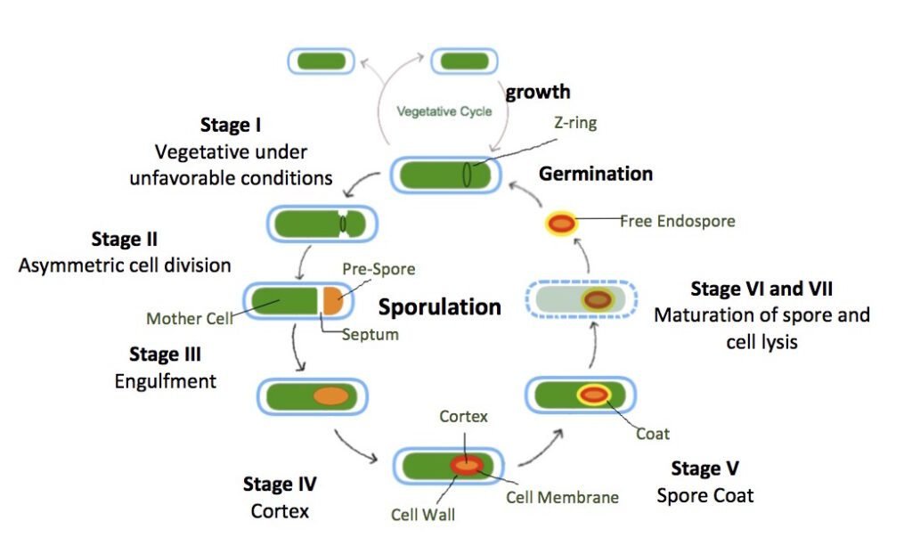
Endospores
Bacteria from the genera Bacillus and Clostridium can form highly resistant internal structures known as endospores, or simply spores. Spores form during the normal life cycle when the environment becomes harsh.
Each vegetative (multiplying) cell generates one spore. Spores can withstand extreme temperatures, extended periods of drying, and other harsh conditions. When the conditions are right, the spore germinates and produces a new vegetative cell, which multiplies and reforms the colony. Spore-forming agents include anthrax, tetanus, botulism, and gas gangrene. Spores contain dipicolinic acid and calcium ions, both of which aid in their resistance.
Source: Wikimedia
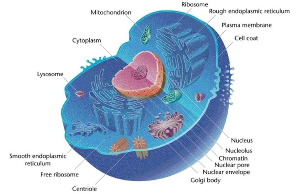
Eukaryotic cell
Eukaryotic cells are those that have both a true nucleus and membrane-bound organelles. Eukaryotes can be unicellular or multicellular. Eukaryotic cells are typically larger and more complex than prokaryotic cells.
They also contain various cellular bodies known as organelles. Organelles play a role in cell activities and serve as compartments for the localization of metabolic function. Microprotozoa, unicellular algae, and fungi all have eukaryotic cells.
Source: Wikimedia
Nucleus
Eukaryotic cells have a distinct nucleus made up primarily of protein and deoxyribonucleic acid (DNA). The DNA is organized into linear units known as chromosomes, or chromatin when the linear units are not visible. Genes are functional chromosomal segments.
Nuclear proteins are classified as histones. Histones act as a supportive framework for DNA in chromosomes. DNA replicates in eukaryotic cells during mitosis.
The nucleus of eukaryotic cells is surrounded by an outer membrane known as the nuclear envelope, which is a double-membrane structure composed of two lipid layers similar to the cell membrane. The nuclear membrane contains pores, which allow the internal nuclear environment to communicate with the cell’s cytoplasm.
The nucleus contains two or more dense masses known as nucleoli (singular, nucleolus). The nucleolus is an RNA-rich area where ribosomes are assembled before exiting the nucleus and entering the cytoplasm.
Cellular organelles
Eukaryotic cells’ cytoplasm (also known as the cytosol) contains a number of microscopic bodies known as organelles. These organelles support a variety of cell functions.
Endoplasmic reticulum
Endoplasmic reticulum (ER), a network of membranes that extends throughout the cytoplasm of eukaryotic cells. In some areas, the ER is studded with sub-microscopic bodies known as ribosomes. This type of ER is known as rough ER. In other places, there are no ribosomes and the ER is known as the smooth ER. The endoplasmic reticulum is where the cell’s protein synthesis occurs. Eukaryotic ribosomes are 80S bodies that hold amino acids together to form proteins. Cisternae are the spaces that exist within the ER membranes.
Golgi body or Golgi apparatus
The Golgi body consists of flattened sacs with curled edges. The outermost sac frequently bulges away to form drop-like vesicles known as secretory vesicles. The Golgi body processes and packages the cell’s proteins and lipids before transporting them to their final destination.
Lysosome
Lysosome is derived from Golgi body. It is a circular, drop-like sac of enzymes located in the cytoplasm. These enzymes aid digestion within the cell. They break down the food particles that enter the cell and make the products available to it. Enzymes are also found in a cytoplasmic body known as the peroxisome.
Mitochondria
The organelle where much energy is released in the Eukaryotic cell is mitochondria. The released energy is used to produce adenosine triphosphate (ATP). The mitochondria are known as the “powerhouses of the cells” because they release and store energy.
Vacuoles
Certain protozoa contain a large, fluid-filled, contractile vacuole. The vacuole, which can take up more than 75% of the cell’s interior, is responsible for water elimination. Water pressure within the vacuole may cause the cell to swell.
Cytoskeleton
The cytoskeleton is an interconnected network of fibers, threads, and interwoven molecules that provides structure to the cell. Microtubules, microfilaments, and intermediate filaments are the three main components of the cytoskeleton. Everything is made up of protein subunits.
Flagella and cilia
Many eukaryotic cells have flagella and cilia. Eukaryotic flagella, like prokaryotic flagella, are long, hairy organelles that emerge from the cell. Eukaryotic flagella, like protozoan flagella, whip around and propel the cell. They are made up of nine pairs of microfilaments arranged around a central pair. Cilia are shorter and more abundant than flagella. Moving cells wave in unison and move the cell. Paramecium is a well-known ciliate protozoan.
Cell wall
Many eukaryotes, including fungi, have a cell wall that exists outside of the cell membrane. Fungi’s cell wall contains chitinas, a complex polysaccharide, as well as some cellulose. In contrast, algal cells lack chitin; instead, their cell walls are entirely composed of the polysaccharide cellulose.
Cell walls support eukaryotic cells and help them resist mechanical pressures, giving them a box-like appearance. The cell walls and membranes are not selective devices.
Cell membrane
The eukaryotic cell membrane follows the fluid mosaic model found in prokaryotic membranes. In eukaryotes, the membrane is a dynamic structure that controls the passage of dissolved molecules and particles into and out of the cytoplasm. However, it lacks the enzymes found in prokaryotic cells and does not function in DNA replication.
Materials must be able to pass through the cell membrane in order for prokaryotic and eukaryotic cells’ cytoplasm to communicate with the outside world. There are several mechanisms for movement.
Various mechanisms of movements
Diffusion
Diffusion is the movement of molecules from a high concentration region to a low concentration area. This movement occurs because the molecules are constantly colliding with one another, and the overall movement of the molecules is away from the region of high concentration. Diffusion is the random movement of molecules, and the pathway they take is known as the concentration gradient. Diffusion is the process by which molecules move down the concentration gradient.
Osmosis
Another method of movement across the membrane is osmosis, which is the movement of water from a high concentration region to a low concentration region. Osmosis occurs across a semipermeable membrane, which allows only certain molecules to pass through while keeping others out. Osmosis is a type of diffusion that uses only water.
Facilitated diffusion
A third mechanism for movement across the membrane is facilitated diffusion, which is aided by certain membrane proteins. The proteins allow only certain molecules to pass through the membrane and promote movement from a high concentration to a low concentration.
Active transport
Active transport is the fourth method for passing across the membrane. When active transport occurs, a protein moves a specific material across the membrane from a region of low concentration to one of high concentration. Because this movement occurs against the concentration gradient, energy must be expended, which is typically derived from ATP.
Endocytosis
Endocytosis, a process in which a tiny patch of cell membrane encloses particles or tiny volumes of fluid at or near the cell surface, is the last mechanism for movement across the membrane. Following its descent into the cytoplasm, the membrane enclosure pinches off from the membrane. When the vesicle contains particulate matter, the process is called phagocytosis; when it contains droplets of fluid, the process is called pinocytosis.
Difference between Prokaryotic cell and Eukaryotic cell
Although these two types of cells are very different, they do share some characteristics. For example, both have cell membranes and ribosomes, but the similarities stop there. The differences between prokaryotic and eukaryotic cells are summarized as follows:
| Prokaryotic cell | Eukaryotic cell | |
| Type of cell | Always unicellular | Unicellular and multi-cellular |
| Cell size | Ranges in size from 0.2μm – 2.0μm in diameter | Size ranges from 10μm – 100μm in diameter |
| Cell wall | Usually present; chemically complex | When present, chemically simple |
| Nucleus | Absent. Instead, they have a nucleoid region in the cell | Present |
| Ribosomes | Present. Smaller in size and spherical in shape | Present. Comparatively larger in size and linear in shape |
| DNA arrangement | Circular | Linear |
| Mitochondria | Absent | Present |
| Cytoplasm | Present, but cell organelles absent | Present, cell organelles present
|
| Endoplasmic reticulum
|
Absent | Present |
| Plasmids
|
Present
|
Very rarely found in eukaryotes |
| Lysosome | Lysosomes and centrosomes are absent | Lysosomes and centrosomes are present |
| Cell division | Through binary fission | Through mitosis |
| Flagella | The flagella are smaller in size | The flagella are larger in size |
| Reproduction | Asexual | Both asexual and sexual |
| Example | Bacteria and Archaea | Plant and Animal cell |
Conclusion
In conclusion, all living organisms are classified as either prokaryotes or eukaryotes based on their cellular characteristics. Prokaryotic cells lack nuclei and organelles, while eukaryotic cells contain both. Prokaryotes include bacteria and cyanobacteria, while eukaryotes include fungi, protozoa, and simple algae. Prokaryotic cells have unique characteristics such as various shapes and sizes, cell walls, and external structures like flagella and pili. Eukaryotic cells are larger and more complex, with organelles such as the nucleus, endoplasmic reticulum, Golgi body, lysosomes, mitochondria, vacuoles, and cytoskeleton. Both prokaryotic and eukaryotic cells have cell membranes that allow for the movement of molecules through diffusion and osmosis.
Frequently asked questions
What is a Prokaryotic cell?
Prokaryotic cells are primitive types of cells that lack a nucleus. Furthermore, prokaryotes lack membrane-bound cellular organelles. Prokaryotes are exclusively unicellular.
What is Eukaryotic cell?
Eukaryotic cells are those that have both a true nucleus and membrane-bound organelles. Eukaryotes can be unicellular or multicellular.
What is the difference between Prokaryotic and Eukaryotic cells?
The nucleus is the defining feature that distinguishes prokaryotic cells from eukaryotic cells. Prokaryotic cells lack a true nucleus, and membrane-bound organelles are found only in eukaryotic cells. Another major distinction between prokaryotic and eukaryotic cells is that prokaryotic cells are all unicellular, whereas eukaryotic cells are not.
Which cell organelle is called powerhouses of cell?
Mitochondrion is called as the powerhouses of cell because they release and store energy.
For more regular updates you can visit our social media accounts,
Instagram: Follow us
Facebook: Follow us
WhatsApp: Join us
Telegram: Join us