Chest XRCT (X-ray Computed Tomography): A Complete Guide
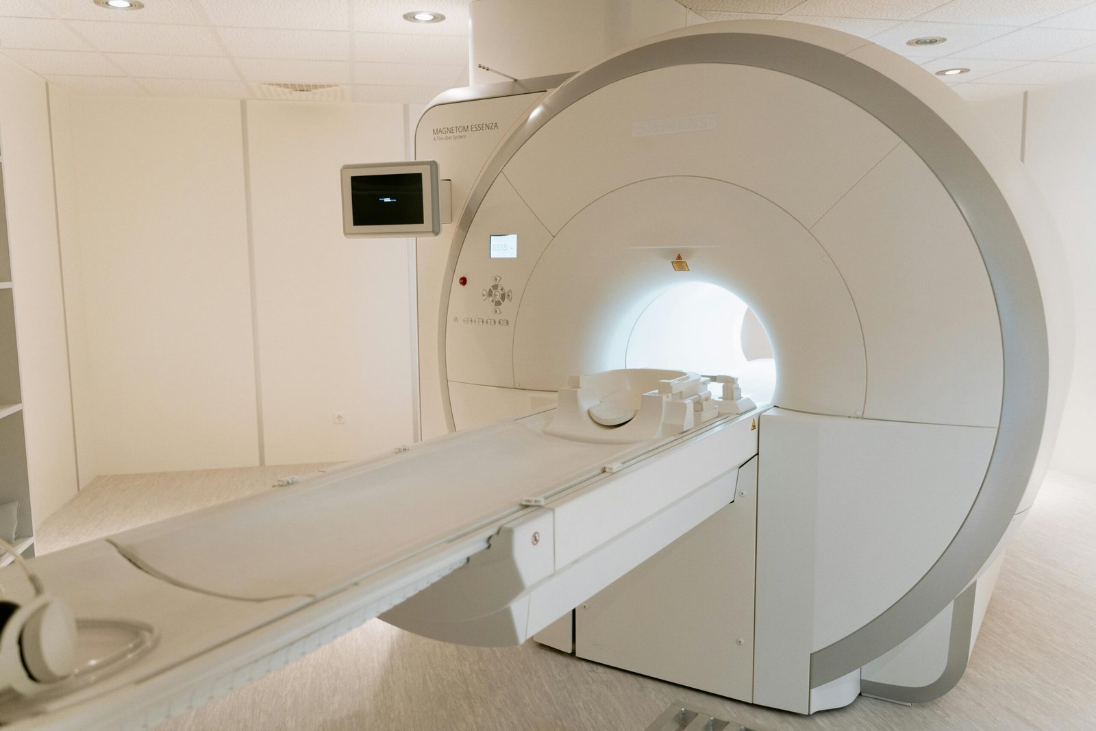
Chest XRCT, also known as Chest CT (Computed Tomography) or Chest HRCT (High-Resolution Computed Tomography), is an advanced imaging technique that combines X-ray images taken from different angles to create detailed cross-sectional views of the chest, including the lungs, airways, heart, blood vessels, and bones of the thoracic cavity.
It plays a crucial role in diagnosing, monitoring, and evaluating a wide range of respiratory and cardiac conditions. Let’s dive deeper into the procedure, preparations, clinical relevance, and safety aspects of this vital diagnostic tool.
What is Chest XRCT?
Chest XRCT is a non-invasive diagnostic test that uses a computer to assemble multiple X-ray images of the chest into 3D or cross-sectional views. It provides far more detail than a regular chest X-ray.
It is especially helpful for evaluating:
- Lung diseases (e.g., pneumonia, tuberculosis, COVID-19, interstitial lung disease)
- Tumors and cancer metastases
- Pulmonary embolism
- Chest injuries
- Structural abnormalities
Types of Chest CT Scans
- Standard Chest CT – Provides an overview of the entire chest.
- HRCT (High-Resolution CT) – Focuses on finer details of lung tissue.
- CT Pulmonary Angiography (CTPA) – Used to detect blood clots in the lungs.
- Contrast-enhanced CT – Uses a contrast dye to better visualize blood vessels or tumors.
Procedure: How Chest XRCT is Performed
Step-by-Step Process:
- Patient Positioning:
- You will lie flat on your back (or sometimes stomach or side) on a motorized CT table.
- Arms may be positioned above the head to avoid interference with the image.
- Preparation for Scan:
- If contrast dye is required, it may be injected through a vein in the arm.
- You may be asked to hold your breath for a few seconds during the scan to reduce motion blur.
- Scanning:
- The CT scanner rotates around the chest, taking multiple X-ray images in slices.
- You may hear a whirring or clicking sound, which is normal.
- The scan usually takes 5 to 10 minutes.
- Post-scan:
- If contrast was used, you’ll be observed for any allergic reaction for a short time.
- Normal activities can typically be resumed immediately.
Preparation Before the Test
1. General Instructions:
- Fasting: If contrast dye is used, fasting for 4–6 hours is usually recommended.
- Clothing: Wear comfortable, loose-fitting clothes. You may be asked to wear a hospital gown.
- Remove Metal Items: Jewelry, glasses, dentures, and underwire bras must be removed as they can interfere with imaging.
2. Inform Your Doctor If:
- You have kidney disease, as contrast dye can affect kidney function.
- You are allergic to iodine or contrast dye.
- You are pregnant or suspect pregnancy (see detailed section below).
- You have diabetes and are on metformin (medication may need to be paused).
Clinical Importance of Chest XRCT
Chest XRCT offers detailed anatomical information, making it vital for accurate diagnosis and treatment planning. Here’s how it’s used in various clinical scenarios:
1. Lung Diseases
- Pneumonia: Identifies extent and complications.
- Tuberculosis: Detects active or healed lesions.
- COVID-19: Assesses severity through ground-glass opacities and fibrosis.
- Interstitial Lung Disease (ILD): HRCT helps differentiate types of ILD.
- Chronic Obstructive Pulmonary Disease (COPD): Evaluates air trapping and bronchial wall thickening.
2. Tumors and Cancer
- Detects lung nodules, masses, or metastases.
- Assesses the spread of cancer to lymph nodes or other structures.
3. Vascular Diseases
- Pulmonary embolism: Detected via CT Pulmonary Angiography (CTPA).
- Aneurysms or aortic dissection: Contrast-enhanced CT can visualize blood vessels precisely.
4. Cardiac Assessment
- Evaluates the heart, pericardium, and coronary arteries in some cases.
5. Trauma and Injury
- Identifies rib fractures, lung contusions, pneumothorax, or hemothorax.
6. Preoperative and Postoperative Monitoring
- Assists in planning lung surgery or evaluating post-surgical complications.
Use of Chest XRCT During Pregnancy
Is it Safe?
Chest XRCT involves ionizing radiation, which poses potential risks to a developing fetus, especially during the first trimester. However:
- The radiation exposure from a single chest CT is relatively low and mostly directed away from the abdomen.
- Shielding (e.g., abdominal lead aprons) is used to protect the fetus.
- It is only performed if absolutely necessary and if the benefits outweigh the risks.
Alternatives During Pregnancy
- Ultrasound or MRI may be preferred, depending on the clinical need.
- Physicians may opt for a chest X-ray with shielding in mild cases.
Risks and Limitations
Risks:
- Radiation exposure (though minimal, it’s cumulative over time).
- Allergic reaction to contrast dye (rare but possible).
- Nephrotoxicity in patients with kidney impairment when contrast is used.
Limitations:
- May not provide functional information like spirometry or PET scans.
- False positives can occur with nodules, requiring biopsy or follow-up imaging.
After the Scan: What to Expect
- Most people can return to normal routines immediately.
- Drink plenty of fluids to help flush out any contrast dye.
- Results are usually available within 24–48 hours and should be reviewed with your doctor.
Tips for Patients
- Don’t panic if you’re sent for a CT scan—it’s a standard and safe diagnostic tool.
- Be honest about pregnancy, allergies, and medical history before the scan.
- If claustrophobic, inform your doctor—you may be given a mild sedative.
- Always follow the pre-scan instructions for accurate results.
Conclusion
Chest XRCT is a powerful, precise, and non-invasive imaging tool that plays a central role in diagnosing and managing various chest conditions. It’s particularly valuable for detecting early disease, guiding treatment decisions, and monitoring response to therapy.
Although it uses a small dose of radiation, the benefits usually outweigh the risks, especially when used judiciously. With the right preparation and awareness, patients can undergo the procedure with confidence and peace of mind.
Frequently Asked Questions (FAQ)
Q1: Is chest XRCT painful?
No. It is a completely painless procedure.
Q2: Can I eat before a chest CT scan?
Yes, unless contrast dye is used—in that case, fasting for 4–6 hours may be required.
Q3: How long does the scan take?
The scanning itself takes about 5–10 minutes.
Q4: Can a chest CT detect cancer?
Yes. It can detect lung tumors, metastases, and lymph node involvement.
Q5: Is it safe during pregnancy?
It is avoided unless critically necessary. Alternatives like ultrasound or MRI are preferred during pregnancy.
For more regular updates you can visit our social media accounts,
Instagram: Follow us
Facebook: Follow us
WhatsApp: Join us
Telegram: Join us